cap


Please see the JBrowse view of Dmel\Cpn for information on other features
To submit a correction to a gene model please use the Contact FlyBase form
AlphaFold produces a per-residue confidence score (pLDDT) between 0 and 100. Some regions with low pLDDT may be unstructured in isolation.
Annotated transcripts do not represent all supported alternative splices within 5' UTR.
Low-frequency RNA-Seq exon junction(s) not annotated.
Gene model reviewed during 5.49
3.0 (northern blot)
3 (northern blot)
None of the polypeptides share 100% sequence identity.
873 (aa); 85 (kD)
865 (aa); 85 (kD)
Homodimer.
Click to get a list of regulatory features (enhancers, TFBS, etc.) and gene disruptions (point mutations, indels, etc.) within or overlapping Dmel\Cpn using the Feature Mapper tool.
The testis specificity index was calculated from modENCODE tissue expression data by Vedelek et al., 2018 to indicate the degree of testis enrichment compared to other tissues. Scores range from -2.52 (underrepresented) to 5.2 (very high testis bias).
Cpn protein is expressed very early in photoreceptor cell development and its expression is not altered in gl eye discs. It is expressed in larvae and adults in photoreceptor cell bodies of the retina. It is also expressed along their axons as they enter the optic lobes. It is also found in ocellar photoreceptors.
Cpn protein is detected in all photoreceptor cells and in ocelli. The staining extends along the length of the photoreceptor cells and along their axons entering the optic lobes.
JBrowse - Visual display of RNA-Seq signals
View Dmel\Cpn in JBrowsePlease Note FlyBase no longer curates genomic clone accessions so this list may not be complete
Please Note This section lists cDNAs and ESTs that fall within the genomic extent of the gene model, which may include cDNAs and ESTs of genes within introns, or of overlapping genes. Please see JBrowse for alignment of the cDNAs and ESTs to the gene model.
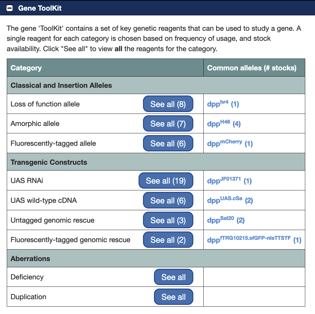
For each fully sequenced cDNA the DGRC maintains various forms of the cDNA (e.g tagged or untagged) in several different host vectors for subsequent cloning and expression in Drosophila and Drosophila cell lines.
monoclonal
Cpn is an immobile Ca[2+] buffer localised along the base of the rhabdomere, which is required to prevent light-induced retinal degeneration.
Class I mutations of Cpn alter the structure of the rhabdomere, class II mutations have rough eyes due to misorientation of the rhabdomeres and photoreceptor cell death. These mutant phenotypes can be rescued by P-element mediated transformation of genomic Cpn DNA. Analysis of the mutants suggests the Cpn plays important roles in both rhadomere development and photoreceptor cell survival.
The Cpn protein binds calcium, contains a long C-terminal leucine zipper and is expressed in the soma and axons of all photoreceptor cells, early in development, when cell-type decisions are being made. Cpn expression pattern is unaffected by mutations in the gl gene, which block photoreceptor development.
Monoclonal antibody 72H5 was used to isolate this protein, which is the same protein as isolated by Martin et al, PNAS 90: 1531--1535, isolated using a different monoclonal antibody, 23E9.
Monoclonal antibody 23E9 used to identify Cpn calcium binding protein in photoreceptor cells and ocelli: the location of the protein within a distinct cytoplasmic region suggests that it might function as a calcium sequestering "sponge" regulating the amount of free cytoplasmic calcium.